Chronic Obstructive Pulmonary Disease is a broad term to define airflow limitation that is not reversible. A patient usually has a component of emphysema, chronic bronchitis or a combination of the two. Diagnosis, analysis and treatment of COPD requires a range of professional medical services; including Medical Imaging, Pathology and Lung Function testing.
Pathology
Arterial Blood Gases – Arterial blood gases are a blood test using blood taken from an artery, most commonly drawn from the wrist. Thie blood test can reveal mild or moderate hypoxemia without hypercapnia in patients with mild COPD. Hypomemia is the partial pressure decrease of oxygen in blood, decreased oxygenation in the blood is a serious condition leading to blacking out, inefficient bodily function and eventually premature death. As the disease progresses, the hypoxemia becomes more severe and hypercapnia develops.
Hypercapnia is where there is a high level of carbon dioxide in the blood, this usually causes a reflex causing increased respiration to increase oxygen and decrease carbon dioxide levels.. Hypercapnia occurs with increasing frequency as the forced expiratory volume in one second (FEV1) falls below one litre, the amount of air exhaled in one second. Blood gas abnormalities worsen during acute exacerbations and may also worsen during exercise and sleep.
Medical Imaging
Chest x-rays and CT scans are imaging studies that are commonly performed in patients with COPD; however, neither is required to diagnose COPD.
Chest x-rays
Prominent Hilar Vascular Shadows
A C.T. Scan showing Lung Bullae
Chest x-rays — Plain chest x-rays have poor sensitivity for detecting COPD. COPD is identifiable on chest x-rays in only around 50% of patients with COPD. Some features on x-rays that can be found (often in advanced disease) include:
Rapidly tapering vascular shadows, increased radiolucency of the lung, a flat diaphragm, and a long, narrow heart shadow on a frontal radiograph, accompanied by a flat diaphragmatic contour and an increased retrosternal airspace on a lateral radiograph. These findings are due to hyperinflation.
Bullae, defined as radiolucent areas larger than one centimeter in diameter and surrounded by arcuate hairline shadows. They are due to locally severe disease, and may or may not be accompanied by widespread emphysema.
Prominent hilar vascular shadows and encroachment of the heart shadow on the retrosternal space. The cardiac enlargement may become evident only on comparison with previous chest radiographs. These findings are due to pulmonary hypertension and cor pulmonale, which can be secondary to COPD.
Computerised tomography — Emphysema can often be diagnosed through computed tomography (CT), but not chronic bronchitis or asthma. High resolution CT has an even greater sensitivity and specificity for emphysema diagnosis than normal CT or plain chest x-ray. CT can be used to determine the location and type of emphysema (centriacinar or panacinar). CT plays an important role in evaluating emphysematous patients for lung volume reduction surgery.
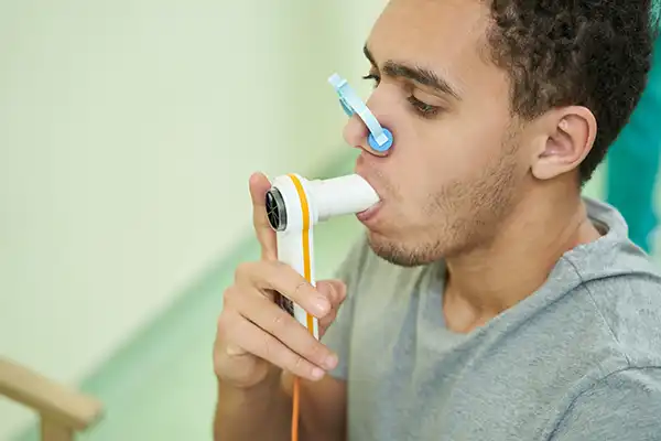
Spirometry is the “Gold Standard” in diagnostic testing of COPD.
Spirometry is a very simple test and measure inhalation and exhalation volumes of a patients breath. In COPD FEV1/FVC ratio should be below 0.7.
This figure is determined from a vast number of studies which have tested lung function in healthy patient and then compared the results to patients confirmed to have COPD. The studies will compare data on sexes, ages and various other demographic variables to identify a range of “normal” functioning.
The patient with COPD has reduced peak expiratory flow, and severely decreased flows at 25%, 50% and 75% of vital capacity compared with the normal range (vertical bars), and shows minimal response to bronchodilator (BD).
By comparison, the patient with chronic asthma shows incomplete, but substantial, reversibility of expiratory flow limitation across the range of vital capacity.
After BD the forced expiratory volume in one second (FEV1) was within the normal range (82% predicted). Absolute and per cent predicted values for FEV1 and forced vital capacity (FVC) before and after BD are shown for each patient.
The Wesley Lung Function Laboratory offers state-of-the-art lung function testing, including spirometry. More information on the Lab, lung function testing, lung conditions and our services can be found HERE.