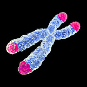
03 Oct Interaction between Obstructive Sleep Apnoea and Shortened Telomere Length on Brain White Matter Abnormality
Previously, magnetic resonance imaging (MRI) has shown an association with severe obstructive sleep apnoea (OSA) and structural brain changes. Within the middle to older age group, 40-80 years, OSA is a common type of sleeping disorder. As a potential biomarker of ageing, telomere length (TL), or shortened TL, has been associated with white brain matter hyperintensities and OSA. Oxidative stress may be enhanced by OSA due to the reduction of antioxidant capacity of blood circulation during nocturnal hypoxia. This oxidative stress may contribute to shorter telomeres, particularly among aged populations.
This cross-sectional study of 420 participants examined obstructive sleep apnoea (OSA) and the interaction between brain white matter changes (WMC) and short TL. They used MRI to determine the status of brain WMC, polysomnography was performed and OSA was determined based on the apnoea-hypopnea index (AHI), and leukocyte TL was measured. The severity of OSA was based on the AHI categorised as normal (<5), mild (5-14.9), moderate (15-29.9), or severe (30). Excessive daytime sleepiness was identified using the Epworth Sleepiness Scale. Quantitative real-time polymerase chain reaction (qRT-PCR) was used to determine TL (measurements n+2,314 defined short). They compared the brain white matter abnormalities with various levels of OSA severities with normal participants.
As displayed in the table above the interaction effect of OSA and short TL was significant when compared to those with neither OSA nor short TL. This study provided first evidence of significant brain WMC abnormalities with mediated interaction of short TL for participants with OSA. As oxidative stress may be related to hypoxia, the mechanisms of impaired brain health in sleep apnoea require further investigation.
Kyung-Mee Choi, PhD; Robert J. Thomas, MD; Dai Wui Yoon, PhD; Seung Ku Lee, PhD; Inkyung Baik, PhD; Chol Shin, MD.


